Publications
 Partners


The lungs are the organs most commonly affected by Covid-19, the disease caused by SARS-CoV-2, and researchers from the UNC School of Medicine have published striking images of cells infected by the virus.
The high-powered microscopic images generated by Dr Camille Ehre from UNC (done in collaboration with two other researchers), show high SARS-CoV-2 viral loads on human respiratory surfaces, ready to spread infection in infected individuals and transmit infection to others.
The images were published in the New England Journal of Medicine.
How it was done
In a laboratory setting, the researchers inoculated the SARS-CoV-2 virus into human bronchial epithelial cells. Using scanning electron microscopy, they examined the cells 96 hours later.
The images below were re-colourised by UNC medical student Cameron Morrison and indicate the following: the infected ciliated cells with strands of mucus (yellow), attached to cilia tips (blue).
In their published paper, the authors explain that cilia are hair-like structures on the surface of airway epithelial cells that transport mucus (and trapped viruses) from the lungs. The airway epithelium’s job is to moisten and protect the airways.
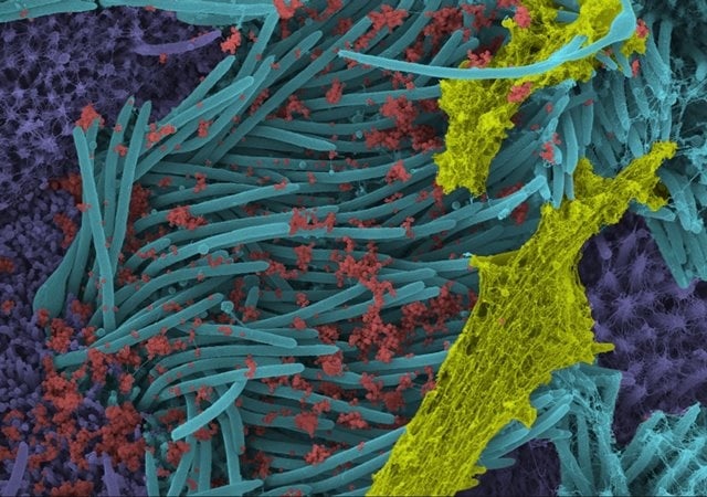
Image source: Ehre Lab, UNC School of Medicine
The image below shows a greater magnification of the structure and density of SARS-CoV-2 virions (red) produced by human airway epithelia. Virions, they wrote, are "the complete, infectious form of the virus released onto respiratory surfaces by infected host cells".
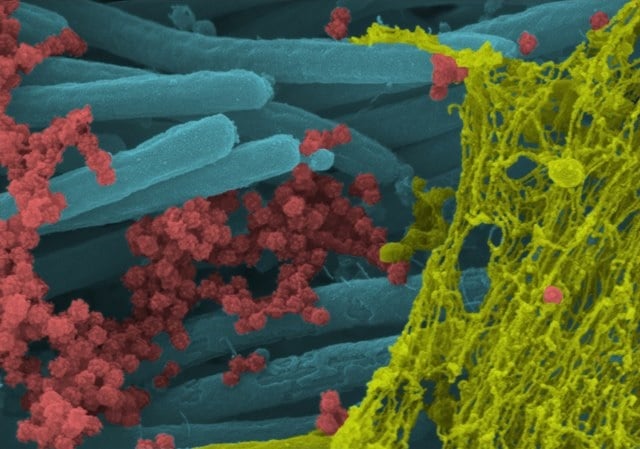
Image source: Ehre Lab, UNC School of Medicine
With the capturing of these graphic images, Ehre wanted to illustrate how intense viral infection of the airways can be.
“This imaging research helps illustrate the incredibly high number of virions produced and released per cell inside the human respiratory system,” a news release by the UNC Health and UNC School of Medicine reads.
According to the researchers, these images make a strong case for the use of face masks in limiting transmission of the virus.