Publications
 Partners


Summary
• Heart valve disease refers to any condition affecting the function of any or all of the four valves of the heart. The valves are located in specific positions, and each valve has a specific function.
• Infection, ischeamic heart disease and congenital abnormalities are important causes of valve disorders.
• Two serious possible complications are bacterial endocarditis and heart failure.
• The initial treatment is often medical, but the definitive treatment is surgical.
What is heart-valve disease?
Heart-valve disease (HVD) refers to any condition disrupting the normal function of the heart valves.
Where are the heart valves located?
There are four valves in the heart: two are inlet valves, two are outlet valves.
Valves are special tissue leaflets located between the heart chambers, permitting blood flow in one direction only. Their position allows them to function as inlet and outlet valves for the important pumping chambers (the left and right ventricles) of the heart. Inlet and outlet valves have a slightly different structure, but their function is the same, i.e. to permit flow in one direction only.
All valves consist of a ring-shaped structure, with the leaflets attached to the inside surface. These open and close. The outlet valves have three halfmoon-shaped leaflets.
Inlet valves have leaflets shaped more like the sails of a boat, and these are attached to specialised heart muscles inside the ventricle, called papillary muscles. These act as "guy ropes" to help stabilise the leaflets against the high pressures within the ventricles.
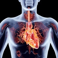
Heart anatomy
The heart is a double pump. One side (the right-hand side) receives used blood from the body, and pumps it on to the lungs where oxygen is replenished. This oxygen-rich blood then returns from the lungs to the left side of the heart, which in turn pumps it out to supply the whole body.
Once body tissues have extracted the oxygen from the blood, the oxygen–depleted blood is returned to the right side of the heart again, and the cycle is repeated.
How do they work?
Tricuspid valve
This tricuspid valve (TV) is found between the right atrium (RA) and right ventricle (RV), and regulates the inflow of "used" blood from the body returning to the heart.
Blood enters the RA and passes through the open TV to the RV. When the RV is full, it contracts to pump the blood onwards to the lungs, where oxygen is replenished. With this contraction, the inlet TV closes so that none of the blood in the RV can squirt back into the RA.
In this way, all the blood is pumped forwards through the pulmonary valve into the lungs.
Pulmonary valve (PV)
The pulmonary valve (PV) is the outlet valve of the RV. This valve is open when the RV contracts (when the TV is closed) so that the blood can only be pumped forwards to the lungs.
The RV thus has an inlet and an outlet valve, controlling the blood flow through the RV. When the RV is relaxed and filling, the inlet valve (TV) is open, and the outlet valve (PV) is closed. When the RV contracts to eject its blood to the lungs, the TV closes and the PV opens.
This occurs with each contraction, ensuring that blood flows in one direction only - that is, onwards towards the lungs.
Mitral valve (MV)
This is the inlet valve for the left ventricle (LV), and is situated between the left atrium (LA), which receives the oxygen-enriched blood returning from the lungs, and the LV which is the main pumping chamber of the heart. A fully open MV allows the LV to fill quickly. As the LV contracts to eject its blood, the MV must close to prevent blood leaking back into the lungs.
Aortic valve (AV)
This is the most important outlet valve – from the LV to the aorta.
It opens when the LV pumps its blood out to the rest of the body. When the LV has emptied, and relaxes (with the MV open) to receive its next load of blood for pumping, the AV closes, preventing any of the oxygenated blood from flowing backwards into the heart again.
The LV thus also has an inlet (mitral) and an outlet (aortic) valve, permitting blood flow in one direction only – that is, from the lungs, forwards into the rest of the body.
What problems can arise?
Mechanically, there are three basic possibilities:
• The valve does not close properly, and thus leaks – also called incompetence, regurgitation or insufficiency.
• The valve does not open properly – called stenosis – impeding filling or emptying of the ventricle.
• A combination of the above two conditions.
An important problem is that of infective endocarditis (IE). This is caused by a bacterial infection of an abnormal valve. The patient becomes extremely ill, and may even need emergency surgery to remove the diseased valve if the infection cannot be controlled by large doses of intravenous antibiotics. The infected valve usually starts to leak quite suddenly.
The condition can be prevented by appropriate use of prophylactic antibiotics. (See the section on "Outcome".)
Reviewed by Dr A.G. Hall (B.Soc.Sc.(SW), MB,Ch.B)
December 2008