Publications
 Partners


• Ultrasound scrotum
• Serum tumour markers: Alphafetoprotein (AFP) and human chorionic gonadotropin (HCG)
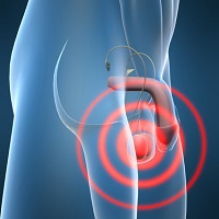
Ultrasound of the scrotum is an excellent test to define the site and nature of scrotal masses. Almost all solid masses of the testes itself are cancerous. Almost all scrotal masses not arising from the testes are benign.
Tumour markers are substances produced by tumours, which can be measured in the blood, thereby indicating the presence and extent of a tumour. Most non-seminomatous germ cell tumours produce AFP and/or HCG. 5-10% of patients with pure seminoma produce elevated levels of HCG. Tumour markers are useful in diagnosis, staging and monitoring of response to treatment. The initial tumour marker levels also provide important prognostic information that may impact on choice of further treatment.
Excision of testicle by radical orchidectomy (see treatment)
The diagnosis of testicular cancer is confirmed by removing the testicle via an incision in the groin and sending the specimen for histological analysis. After the diagnosis has been confirmed, the next steps are a series of staging investigations to determine the presence and extent of spread of disease.
Staging Investigations
•Chest X-ray
•CT scan of abdomen and pelvis
•Tumour markers (HCG and AFP)
The subsequent management following orchidectomy will depend on the nature of the primary tumour and the stage (extent) of spread of disease. The CT scan is performed to detect spread to the lymph glands around the aorta and to identify liver metastases.
A chest X-ray will detect 90% of lung metastases. If the tumour marker levels were elevated prior to removal of the testicle they are repeated. Tumour markers which remain elevated after removal of the testis indicate the presence of residual disease.
Staging
A number of staging systems are in use throughout the world. This complicates the comparison of data from different sentra. The Royal Marsden Hospital system is widely used and is simple and easy to understand. It is also widely used in South Africa.
• Stage I - Tumour confined to the testes
• Stage II - Tumour spread to para-aortic lymph glands
• Stage IIa - Glands less than 2cm
• Stage IIb - Glands 2-5cm
• Stage IIc - Glands more than 5cm
• Stage III - Lymph gland involvement in chest or neck
• Stage IV - Spread outside of lymph glands, i.e. to lung, liver, bone or brain
Previously reviewed by Dr Pieter J le Roux MBChB, FRCS(Eng), FRCSI, FCS(SA)Urol.
Reviewed by Dr David Eedes, Oncologist, February 2011