Publications
 Partners


Alternate names
Upper gastro-intestinal tract (GIT) study
Contrasted swallow
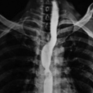
What is a barium swallow?
This is an investigation that allows visualisation of the oesophagus during the swallowing process. Under normal circumstances the oesophagus cannot be seen clearly during swallowing on routine radiographs. With the aid of repetitive x-rays (fluoroscopy) and a contrast agent named barium, the swallowing mechanism and oesophagus can be accurately examined. This enables the radiologist to diagnose several diseases that affect the oesophagus.
Common uses
This investigation is frequently performed in the evaluation of patients who complain of heartburn, upper abdominal pain, painful swallowing or even the inability to swallow liquid or solid food. It is useful in the diagnosis of oesophageal cancer, reflux disease, hernias and perforation of the oesophagus.
It can be used to demonstrate the exact location and severity of diseases of the oesophagus. Treatment of these oesophageal diseases can be planned and response to treatment can be followed with this investigation.
How should I prepare?
Patients should be starved from the night prior to the investigation (in other words be kept nil by mouth from 10pm on the evening before the examination) until after the examination. No sedation or anaesthetic is used during the investigation, thus allowing the patient to be fully awake and able to co-operate during the investigation. Therefore, patients will be able to drive themselves to and from the investigation and be able to presume their normal daily activities directly afterwards.
How is the procedure performed?
In the radiology department, it will be explained to the patient exactly what is expected of them. The procedure will be overseen by the radiologist and a radiographer. While standing, the patient will be positioned in front of the x-ray machine. The x-ray machine will be focused on the neck and chest. The patient will have to swallow the barium or other contrast agent on demand. The contrast agent will coat the lining of the oesophagus thus making it easy to see the oesophagus on the images created by the x-ray machine during swallowing. These images are then evaluated by the radiologist in order to make a diagnosis.
When the patient has severe swallowing difficulties and some of the barium leaks into the trachea, the patient will aspirate causing the contrast to enter the lungs. This usually cause coughing and discomfort.
The exposure to x-rays always carry a theoretical risk of developing certain cancers, but the chances of this is extremely low seeing that the exposure to x-rays is limited and the examination performed under strict supervision.
Limitations
Although this investigation is very accurate in demonstrating diseases of the oesophagus, a specific diagnosis cannot always be made. In some cases, a biopsy of the affected area is needed to make a diagnosis.