


 Publications
 Partners


Alternate names
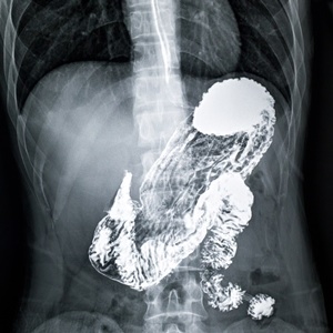
Upper gastro-intestinal tract seriesBarium swallow and follow through
What is a barium meal?
This is an investigation that allows visualisation of the oesophagus, stomach and first part of the small bowel (duodenum) with the use of X-rays and a contrast medium called barium. Under normal circumstances the stomach cannot be seen clearly on plain radiographs. After swallowing the contrast medium, it coats the lining of the oesophagus, stomach and duodenum allowing visualisation of these organs on radiographs. Diseases affecting the stomach can then be accurately diagnosed.Common uses
The study is commonly performed in patients who complain of heartburn, unexplained weight loss, abdominal pain, blood in the stool (melena) or reflux disease.It is used to diagnose stomach/peptic ulcer disease, gastro-oesophageal reflux disease, stomach cancer, stomach outlet obstruction, hiatus hernias and several other diseases.
How should I prepare?
The patient should be starved from the night before the examination and not eat or drink anything on the day of the investigation. No sedation or anaesthetic is used during the investigation, so the patient can drive themselves to and from and presume their normal daily activities.How is the procedure performed?
The entire procedure will be explained by the radiologist on the day of the investigation. Before the swallowing of the barium, the patient will be injected with a medication that will slow down the normal contractions of the stomach. Gas-producing tablets are also given. These will distend the stomach, allowing it to be better coated by the contrast agent.Once the barium is swallowed, the patient is turned around on the x-ray table to ensure that the entire stomach is well coated with the contrast material. X-rays will then be taken in various positions. The lining of the stomach and duodenum are then evaluated from these images.
Risks
Barium has a chalky taste and may cause some constipation a few days after the investigation.The exposure to x-rays always carry a theoretical risk of developing certain cancers, but the chances of this is extremely low seeing that the exposure to x-rays is limited and the examination performed under strict supervision.
Limitations
This study allows excellent visualisation of the lining of the stomach and duodenum, but cannot always diagnose the disease accurately. In these cases a gastroscopy might be indicated.


