Publications
 Partners


How is the exam done?
The doctor may first spray the nose with a type of local anaesthetic before inserting a flexible rubber or rigid steel tube called an endoscope to look inside of the patient’s nose. Although this process is not painful, the patient may experience slight discomfort.
Physical findings:
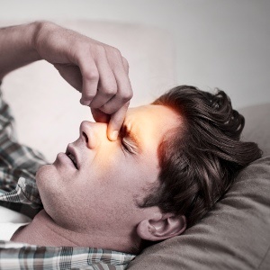
Physical findings may include redness and swelling of the nasal passages, purulent (pus-like) drainage, tenderness to percussion (tapping) over the cheeks or forehead region, and swelling around eyes and cheeks.
Diagnoses further explained:
An X-ray may be done to confirm whether mucus is present in the sinuses. Depending on what is found at the time of the endoscopy, a computerised axial tomography (CAT) scan of the sinuses may be needed. If surgery is needed, a CAT scan will ordinarily be done prior to surgery. CAT scans and magnetic resonance imaging (MRI) scans are much more sensitive in their ability to diagnose sinusitis, but are expensive.
What is a rhinoscopy?
This is a procedure for looking into the back of the nasal passages with a small flexible fibre-optic tube, which may be used to look directly at the sinus openings and check for obstruction. It may sometimes be necessary to perform a needle aspiration (drawing of fluid) of a sinus to confirm the diagnosis of sinusitis, and to collect infected material. This will be cultured so the bacteria causing the infection may be identified.
Read more:
Treating sinusitis
Reviewed by Dr Harris Steinman MBChB. (UCT), D.CH (SA), FAAAAI, D.Av.Med.(SAMS), Private specialist at FACTS (Food & Allergy Consulting & Testing Services), February 2015. (Previously updated by Dr H Steinman, June 2007)