Publications
 Partners

Sorry, this category has no content yet.


Rand - Dollar
19.09
+0.1%
Rand - Pound
23.78
+0.0%
Rand - Euro
20.44
+0.0%
Rand - Aus dollar
12.45
-0.4%
Rand - Yen
0.12
+0.1%
Platinum
925.70
+0.6%
Palladium
1,036.00
+1.0%
Gold
2,327.99
+0.3%
Silver
27.45
+0.5%
Brent Crude
88.42
+1.6%
Top 40
68,051
+0.8%
All Share
74,011
+0.6%
Resource 10
59,613
-2.2%
Industrial 25
102,806
+1.7%
Financial 15
15,897
+1.8%
All JSE data delayed by at least 15 minutes

WATCH | From delivery driver to graduate: Tshireletso Makabe's journey to honour...
2h ago

How a Northern Cape village turned an alien tree into superfood coffee
15 Apr
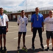
Blades of glory: Mitchells Plain speed skaters bag 9 medals at SA Rollersport...
02 Apr
Cox Thato and her crew pull out all the stops for prestigious UK tour
08 Apr