


 Publications
 Partners

Sorry, this category has no content yet.


Rand - Dollar
18.81
+1.1%
Rand - Pound
23.50
+1.2%
Rand - Euro
20.12
+1.4%
Rand - Aus dollar
12.29
+0.9%
Rand - Yen
0.12
+2.5%
Platinum
923.80
-0.2%
Palladium
959.00
-3.2%
Gold
2,338.80
+0.3%
Silver
27.21
-0.8%
Brent Crude
89.01
+1.1%
Top 40
69,358
+1.3%
All Share
75,371
+1.4%
Resource 10
62,363
+0.4%
Industrial 25
103,903
+1.3%
Financial 15
16,161
+2.2%
All JSE data delayed by at least 15 minutes

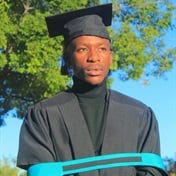
WATCH | From delivery driver to graduate: Tshireletso Makabe's journey to honour...
24 Apr
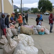
How a Northern Cape village turned an alien tree into superfood coffee
15 Apr
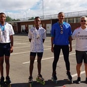
Blades of glory: Mitchells Plain speed skaters bag 9 medals at SA Rollersport...
02 Apr
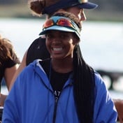
Cox Thato and her crew pull out all the stops for prestigious UK tour
08 Apr
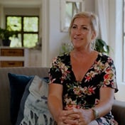
How this work-from-home mom gets back-to-school ready with the help of Mac
10h ago
Watch | Rhodes University Marks 120 Years of Academic and Cultural Milestones
10h ago

Civil Engineering student Zaid’s building blocks to success start with his trusty Mac
25 Apr

Why the iPad is the ultimate study buddy for learners from grades R-12
23 Apr
