


 Publications
 Partners


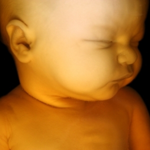
Using real-time images of brain connections developing in late-stage foetuses, scientists say they've been able for the first time to compare the order and strength of these connections.
The research, though very preliminary, might one day lead the way to more effective therapies for brain disorders such as dyslexia, attention-deficit/hyperactivity disorder (ADHD) and autism, the researchers said.
Scientists from the US National Institute of Child Health and Human Development and Wayne State University School of Medicine looked at 25 foetal brains from a group of pregnant women between 24 and 38 weeks of gestation. The researchers used an imaging technique called functional MRI (fMRI) to visualise "communication" between various regions of the brain.
The scientists learned that connections between the right and left sides of the brains got stronger as foetuses grew older. They said they also learned that shorter distances between matching areas produced stronger signals than longer spans between corresponding areas on the brain's outer edges.
"What we're seeing is a picture of emerging connectivity that the right and left side are kind of building a bridge to each other," said study author Moriah Thomason, an assistant professor of paediatrics at Wayne State, in Detroit. "It's evidence for the fact that already in foetal life, this anatomy is being constructed in a way that we would expect. But for the first time, we can show the development of these networks."
Better understanding of conditions
While scientists had previously used fMRI scans on foetuses, this new research is the first to compare a group of foetuses and identify features of development, Thomason said. Nearly 90% of the pregnant mothers participating in the study were black, while the rest were white or multiracial. They later delivered 17 boys and eight girls.
MRI scans don't emit radiation, thus minimising risk to the foetuses during the procedure. The technique showed significant connections between half of the dozens of brain areas tested.
The findings might provide groundwork for understanding how and when brain development may go awry during gestation, Thomason said. This may possibly lead to better understanding of conditions such as ADHD, dyslexia and autism, which are thought to arise from disrupted brain networks, she said.
"If we know what gets in the way of those [normal] processes, we have a better shot at treating those disorders," Thomason said. "It's not just about early identification. An additional valuable outcome is, when you can see what normal looks like and see what disruption looks like, you have the opportunity to pick out patterns that tell you about the origin of that disease," she explained.
"The brain can be a tattletale to what is going on in those diseases," Thomason added, "and that can help us develop novel treatments."
More information
The US National Library of Medicine has more on foetal
development.


