


 Publications
 Partners


Alternative names
Pleuroscopy or video assisted biopsy.
What is a Pleural biopsy and why is it done?
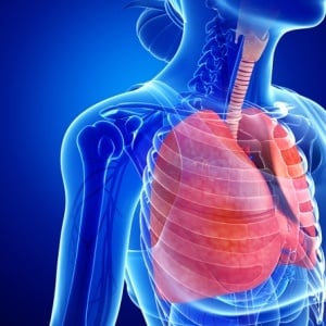
The pleura are the fibrous linings of the lung. There are two different linings - the first, on the surface of the lung itself and the second on the inside surface of the rib cage. These two layers are usually touching with no air or water between them. An accumulation of fluid "water around the lung" (pleural effusion) in this space can be seen on a chest X-Ray. Common causes for pleural effusions are tuberculosis, cancer or trauma. To be absolutely certain of the diagnosis, a small piece of the pleura needs to be removed and examined by a pathologist under a microscope. This is done by a pleural biopsy.
How is it done?
The simplest and quickest way of doing a pleural biopsy is a "closed" biopsy using an Abrams needle (about size of a knitting needle). The skin and muscle are numbed using a local anaesthetic injection after which the needle is inserted into the space between the lung and rib cage (where the water has accumulated). A few small pieces of the pleura (rib side) are then removed. No stitches or pipes drains are usually required.
Alternatively a "keyhole" surgery approach can be used where 2- 3 holes are used and a video camera (scope) is used to look inside the ribs and biopsy the pleura. This technique requires more time and preparation and usually requires an operating theatre. To allow a better view of the lung lining, the lung needs to be collapsed first by allowing air into the space around the lung. One can breathe quite comfortably using the other lung. After the biopsy a pipe drain is left in the space until all the air has come out and the lung has re-expanded. This procedure can be used to biopsy the lung lining as well but does require a pipe drain to be left in for a few hours until all the air and fluid has drained out.
What are the risks?
The major risks involved are pain, bleeding, infection and collapsing of the lung. Because of the nature of the nerve supply to the pleura, a completely painless biopsy is difficult. In good hands, the other risks are minimal. Occasionally, air can leak into the space after a closed biopsy. This usually does not require any treatment unless the lung collapses down or a large amount of air has leaked in. A pipe drain will then be inserted to remove the air.
Do I need to prepare for it?
As the procedure is done as an outpatient, there is usually no preparation required. If a video scope is being used, your doctor may ask you not to eat for a few hours beforehand.
What are the limitations?
The biggest limitation of a pleural biopsy is that the piece removed may not contain enough tissue for the pathologist to make a diagnosis. The video scope although more of a complicated procedure, generally has a better success rate as one can see usually see what to biopsy. The closed biopsy done by an experienced doctor will be sufficient in most cases.
Dr Richard van Zyl-Smit (Specialist Physician and Pulmonologist) MBChB MRCP(UK) FCP(SA) MMED Cert Pulm(SA)


