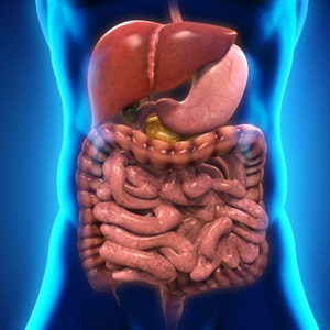
There are three parts to making a correct diagnosis of digestive disorders.
These include a scrutiny of the medical history of the patient, a physical examination, possibly also a psychological evaluation, and doing appropriate tests to obtain tissue samples, images or stool samples from the patient.
Medical history
These would include questions about exact symptoms, general lifestyle choices, diet, previous gastro-intestinal disorders, and any systemic disorders, such as diabetes.
Specific questions can relate to the nature of the pain or discomfort experienced by the patient, where it is located, and whether there are certain actions or foods which make it worse. The doctor will ask about bowel movements and the type of stools produced.
Physical examination
According to the Merck Manuals, a physical examination would entail the following:
• Observation of the abdomen from different angles to see if any swelling is visible
• A stethoscope is used to listen for any sounds emanating from the abdomen
• The doctor feels for tenderness or enlarged organs or abnormal masses
• Pain caused by gentle pressure could indicate inflammation or infection of the lining of the abdominal cavity
Your doctor will also do a rectal examination if indicated. This might be uncomfortable.
A psychological evaluation is sometimes done, as there can be a close connection between digestive problems and psychological conditions such as depression and anxiety. According to the Merck Manuals psychological factors play a role in as many as 50% of people with digestive disorders.
Tests to diagnose digestive disorders
Some tests need the digestive system to be empty, some require no ingestion of food for eight to 12 hours before the test, and others require no preparation. Many of these tests make use of sophisticated equipment and can be very expensive.
Stool tests. This test is used to check for the presence of blood in the stool, or the presence of parasites, fungi, viruses, bacteria, white blood cells, bile, or cancer. It can also be used to check whether a patient is experiencing poor absorption of nutrients.
Endoscopes. An endoscope is a device with a light attached that is used to look inside a body cavity or organ, according to the Medline Plus Medical Encyclopaedia. The scope is inserted through a natural opening such as the mouth or the anus. An endoscope is used to examine the oesophagus (oesophagoscopy), the stomach (gastroscopy), the anus and the rectum (sigmoidoscopy) and the entire large intestine, the rectum and the anus (colonoscopy).
Intubation of the digestive tract. A small flexible tube is passed through the nose or mouth into the stomach or the small intestine. This can be used to diagnose or treat disorders, or to obtain a sample of stomach fluid. These stomach secretions can be tested for the presence of blood, acid levels or enzyme levels can also be tested, according to the Merck Manuals.
Laporoscopy describes an examination of the abdominal cavity using an endoscope that is inserted through a small incision in the navel. This is used to look for tumours or other abnormalities, take tissue samples, examine surrounding organs, and to do surgery.
Paracentesis (Insertion of a needle). This is done to get a sample of fluid from the abdominal cavity or other parts of the digestive system.
Tests for acid and reflux. A small tube is placed in the oesophagus to test acid reflux into the oesophagus.
Different types of imaging techniques
• Normal X-rays. These can show a blockage or a paralysis in the digestive system.
• Barium X-rays. The patient swallows barium before the X-ray is taken. The barium shows up white on the X-rays and can show whether the oesophagus and stomach function normally. Barium can also be used to look for polyps or tumors or structural abnormalities in the lower intestine.
• Computed Tomography Scan (CT scan) and Magnetic Resonance Imaging (MRI). These are used to assess the size and the location of organs in the abdominal cavity. These tests can also detect tumours (both malignant and benign) and changes in the blood vessels. CT scans create two-dimensional and three-dimensional images of the abdominal area, the colon or the small inetsine, making detection of abnormalities easier.
• Ultrasound scanning. This technique uses sound waves to produce images of internal organs and any abnormalities present in them.
Read More:
Preventing digestive disorders
Symptoms of digestive disorders
Reviewed by Dr Estelle Wilken (MBChB) (MMed Int) ,Senior Specialist, Internal Medicine and Gastroenterology, University of Stellenbosch and Tygerberg Hospital. February 2016.


 Publications
 Partners


